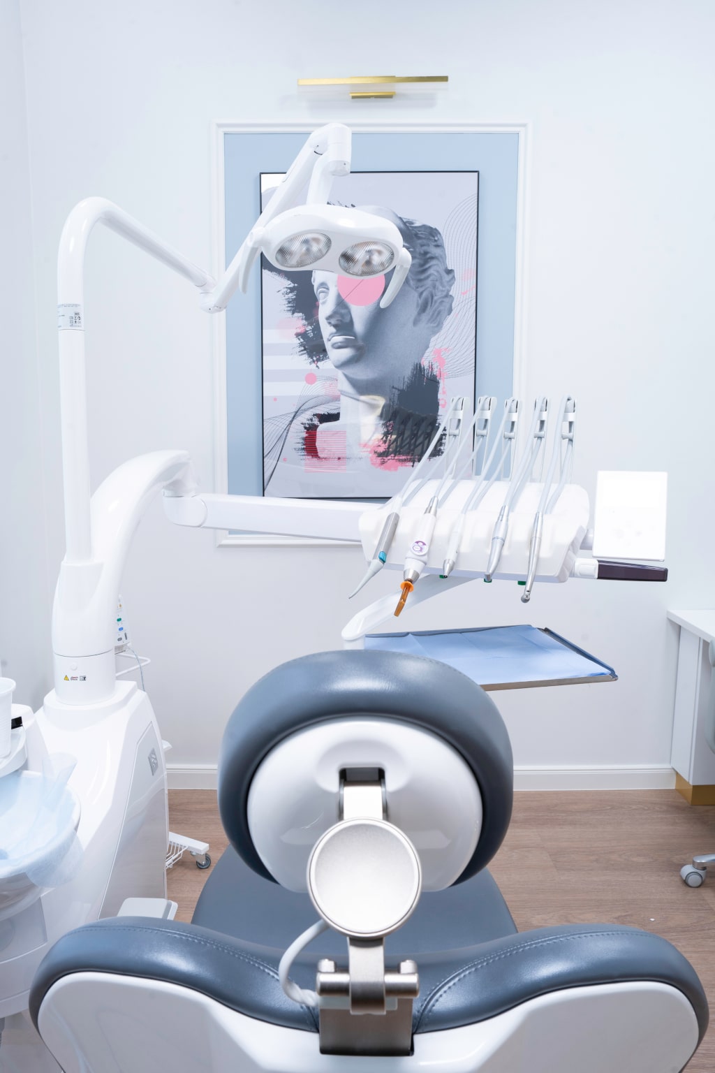
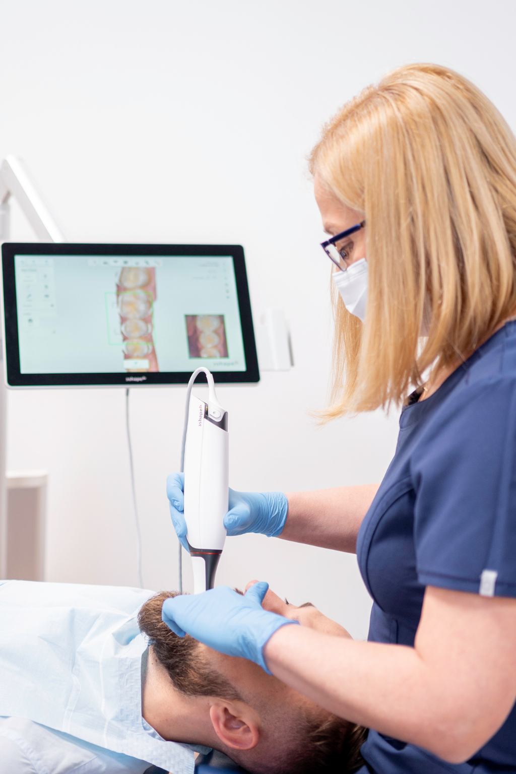
Diagnostics

CBCT computed tomography of the maxilla and mandible
from 299 PLN
CBCT tomography allows you to create a three-dimensional image helpful in assessing the condition of the jaw, teeth, facial bones, nasal cavity and sinuses. In-depth diagnostics is an important element of preparation for the procedure in the field of aesthetic dentistry, it facilitates implantation, surgery and endodontic treatment.
Tomografia CBCT szczęki i żuchwy, czyli inaczej tomografia stożkowa, usprawnia też diagnozowanie chorób przyzębia, zaburzeń słuchu, stanów zapalnych zatok i wielu innych dolegliwości. Mimo że badanie zajmuje dosłownie kilkadziesiąt sekund, to daje możliwość wykrycia chorób na wczesnym etapie i znalezienia źródeł uporczywego bólu.
CBCT of the maxilla and the mandible, or cone tomography, also improves the diagnosis of periodontal diseases, hearing disorders, sinusitis and many other ailments. Although the examination takes literally tens of seconds, it makes it possible to detect diseases at an early stage and find the sources of persistent pain.
3D image indispensable in modern dentistry
CBCT computed tomography can be safely called a revolutionary technique of maxillofacial imaging, because it allowed the use of 3D images in dentistry instead of 2D. During this examination, the X-ray beam takes the shape of a cone, creating numerous views around the patient, which are later used to generate 3D images. Cone tomography is a much more accurate examination than a panoramic image – in the case of a CBCT image, we obtain a 3D model, and a pantomogram gives us a 2D image.
The advantage of this innovative diagnostic method is the simultaneous reduction of the radiation dose and the shortening of the examination time. In this case, the radiation dose was reduced tenfold in relation to traditional tomography. Despite this, the image of the jaw and its surroundings obtained by CBCT tomography is of much higher quality.
Extensive use of three-dimensional images
CBCT has been shown to be effective in detecting and treating jaw tumors, examining bone structure and determining the orientation of the tooth. Thus, such three-dimensional images provide doctors with a lot of information necessary to plan professional tooth reconstruction with the help of implants.
Due to precise measurements, cone tomography turns out to be reliable when planning surgical procedures. Thanks to this technique, it is possible to thoroughly examine the tooth structure and its root canals.
- Quick and comfortable examination – it only takes 10-20 seconds
- Universal use in many areas of dentistry
- Proven effectiveness in the rapid detection of lesions